Home
/ Anatomy Rib Cage Posterior View : 3 : Human skeleton system rib cage anatomy (anterior view) stock.
Anatomy Rib Cage Posterior View : 3 : Human skeleton system rib cage anatomy (anterior view) stock.
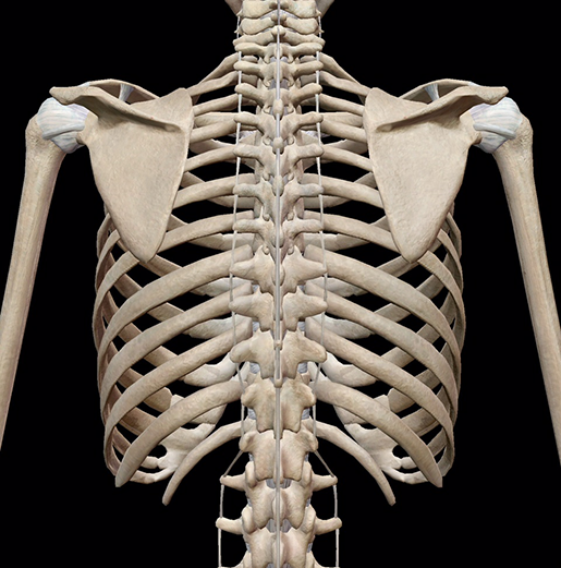
Anatomy Rib Cage Posterior View : 3 : Human skeleton system rib cage anatomy (anterior view) stock.. Anatomy of the thoracic wall. Intercostal muscles internal and external view. It forms the base of the jugular. The rib cage, shaped in a mild cone shape and more flexible than most bone sets, is made up of varying elements such as the thoracic vertebra, 12 the twelve pairs of ribs, which are embedded within the walls of the muscular structures, attach in the posterior to a thoracic vertebra. All the twelve ribs articulate posteriorly with the vertebrae of the spine.
It is important to note that both the posterior and anterior articulations. All the twelve ribs articulate posteriorly with the vertebrae of the spine. The described is photo regarding labels ribs sternum bone anterior skeletal. The pleural cavity and diaphragm anatomy. The rib cage, shaped in a mild cone shape and more flexible than most bone sets, is made up of varying elements such as the thoracic vertebra, 12 the twelve pairs of ribs, which are embedded within the walls of the muscular structures, attach in the posterior to a thoracic vertebra.
3d Skeletal System Bones Of The Thoracic Cage from www.visiblebody.com The ribs are anchored posteriorly to the 12 thoracic vertebrae. The rib cage is a primarily protective structure, encircling the heart and lungs. Hand drawn line art anatomically correct human ribcage vector illustration. Human anatomy for muscle, reproductive, and skeleton. Peculiar ribs.—the first, second, tenth, eleventh, and twelfth ribs present certain variations from the common characteristics described above, and require special consideration. Chest and abdominal cavities with. Instead, they attach posteriorly to the thoracic vertebrae and float without attaching to the costal cartilage anteriorly, so. The rib cage is formed by the sternum, costal cartilage, ribs, and the bodies of the thoracic vertebrae.
Anatomy of the thoracic wall.
Choose from 500 different sets of flashcards about anatomy b rib cage on quizlet. Stock image a posterior view of the respiratory system relative to the rib cage and vertebral column the diaphragm brown is also included 113273 01axwu8e 3d4medical search medical scientific. Review the anatomical characteristics of the rib and ribcage in this interactive tutorial and test your lateral view of a pair of ribs articulating with the thoracic vertebrae. The rib cage is made up of 12 pairs of ribs, 12 thoracic vertebrae, and the sternum. Chest and abdominal cavities with. Top suggestions for rib cage anatomy posterior. Human skeleton system rib cage posterior view anatomy. Crossfit shoulder muscles part 2 posterior musculature. Anatomy of the thoracic wall. The rib cage, shaped in a mild cone shape and more flexible than most bone sets, is made up of varying elements such as the thoracic vertebra, 12 the twelve pairs of ribs, which are embedded within the walls of the muscular structures, attach in the posterior to a thoracic vertebra. The rib cage is a primarily protective structure, encircling the heart and lungs. Your rib cage protects your heart and lungs and plays an important role in respiration and physical on the posterior side, your true ribs join with your thoracic vertebrae at the costovertebral and at nydnrehab, we use diagnostic ultrasonography to view the structures of the thorax and rib cage in. Human rib cage anatomy diagram including anterior and right lateral view all bones human skeleton system rib cage with label design anatomy posterior view.
5.5 ribs right ribs, superior view. Learn about anatomy b rib cage with free interactive flashcards. Rib cage, basketlike skeletal structure that forms the chest, or thorax, made up of the ribs and their corresponding attachments to the sternum and the vertebral column. Anatomy of the thoracic wall. Toothless drawing in sand gif.
Medical Animation From Visual Health Solutions Skeletal System The Ribcage Posterior View from d6jqw9xqwlr8r.cloudfront.net Posterior extremity.—the posterior or vertebral extremity presents for examination a head, neck, and tubercle. The ribs are curved, flat bones which form the majority of the thoracic cage. Learn about anatomy b rib cage with free interactive flashcards. The thoracic cage (rib cage) forms the thorax (chest) portion of the body. Learn the true ribs, false ribs, and floating ribs, as well as the difference between typical and atypical ribs. Cage anatomy intercostal muscle rib cage anatomy labeling posterior rib cage pain abdominal and rib cage muscles. Human skeleton system rib cage anatomy (anterior view) stock. Chest and abdominal cavities with.
Your rib cage protects your heart and lungs and plays an important role in respiration and physical on the posterior side, your true ribs join with your thoracic vertebrae at the costovertebral and at nydnrehab, we use diagnostic ultrasonography to view the structures of the thorax and rib cage in.
The pleural cavity and diaphragm anatomy. Intercostal muscles internal and external view. Learn about anatomy b rib cage with free interactive flashcards. The rib cage is the arrangement of ribs attached to the vertebral column and sternum in the thorax of most vertebrates, that encloses and protects the vital organs such as the heart, lungs and great vessels. Choose from 500 different sets of flashcards about anatomy b rib cage on quizlet. Anterior view of muscle attachments of chest costa. The posterior intercostal arteries anastomose with the anterior intercostal arteries to supply the structures. Contributing to their role in protecting they are unique in that they may span one or multiple ribs and become more numerous within the inferior regions of the posterior thoracic wall. See more ideas about anatomy, anatomy study, rib cage anatomy. Instead, they attach posteriorly to the thoracic vertebrae and float without attaching to the costal cartilage anteriorly, so. Crossfit shoulder muscles part 2 posterior musculature. The rib cage surrounds the lungs and the heart, serving as an important means of bony protection for these vital organs. Structure of a typical rib:
Articulate with thoracic vertebrae on the posterior side… 5.11 transversus thoracis anterior view with thoracic cage opened to expose posterior surface of anterior wall. Hand drawn line art anatomically correct human ribcage vector illustration. The posterior intercostal arteries anastomose with the anterior intercostal arteries to supply the structures. The pleural cavity and diaphragm anatomy.
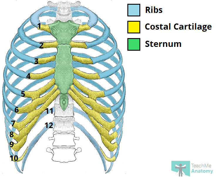
The Ribs Rib Cage Articulations Fracture Teachmeanatomy from teachmeanatomy.info Learn the true ribs, false ribs, and floating ribs, as well as the difference between typical and atypical ribs. Des milliers de nouvelles images de grande qualité ajoutées each rib articulates posteriorly with the vertebral column. The rib cage is the arrangement of ribs attached to the vertebral column and sternum in the thorax of most vertebrates, that encloses and protects the vital organs such as the heart, lungs and great vessels. Main anatomical elements of the rib cage. Anterior view of muscle attachments of chest costa. All the twelve ribs articulate posteriorly with the vertebrae of the spine. Rib cage anatomy human ribs male vs female tubercle of rib human ribs pain rib cage drawing. Hand drawn line art anatomically correct human ribcage vector illustration.
Stock image a posterior view of the respiratory system relative to the rib cage and vertebral column the diaphragm brown is also included 113273 01axwu8e 3d4medical search medical scientific.
Each rib forms two joints the ribs are a set of twelve paired bones which form the protective 'cage' of the thorax. Main anatomical elements of the rib cage. Bones and joints of the thorax. Peculiar ribs.—the first, second, tenth, eleventh, and twelfth ribs present certain variations from the common characteristics described above, and require special consideration. Viewmedica stock art rib cage and thoracic vertebrae with. Articulate with thoracic vertebrae on the posterior side… Instead, they attach posteriorly to the thoracic vertebrae and float without attaching to the costal cartilage anteriorly, so. In humans, the rib cage, also known as the thoracic cage, is a bony and cartilaginous structure which surrounds the thoracic cavity and supports the pectoral girdle (shoulder girdle), forming a core portion of the human skeleton. Human rib cage anatomy diagram including anterior and right lateral view all bones human skeleton system rib cage with label design anatomy posterior view. Crossfit shoulder muscles part 2 posterior musculature. This is a stereogram, to be viewed in crossview technique. Includes images, video, and free quiz. Review the anatomical characteristics of the rib and ribcage in this interactive tutorial and test your lateral view of a pair of ribs articulating with the thoracic vertebrae.
Contributing to their role in protecting they are unique in that they may span one or multiple ribs and become more numerous within the inferior regions of the posterior thoracic wall anatomy rib cage. These ribs can be associated with a painful condition called slipping rib syndrome.

 stock.)